Product Sheet CP10217
Description
BACKGROUND Receptor tyrosine kinase like orphan (Ror) proteins are type-I transmembrane receptor tyrosine kinase members of the neurotrophic tyrosine kinase receptor superfamily. The extracellular region of Ror proteins contains an immunoglobulin domain, a Cys-rich domain, also called the Frizzled domain, and a Kringle domain. Intracellularly, Ror proteins possess a tyrosine kinase domain and a proline-rich domain straddled by two Ser/Thr-rich domains. The vertebrate Ror family members, Ror1 and Ror2, shares 58% amino acid sequence identity. Analyses of mutants that are defective in the single nematode Ror demonstrate a role in cell migration and in orienting cell polarity. Mice lacking one of the two Ror gene products display defects in bone and heart formation. Similarly, two different human bone development disorders, dominant brachydactyly B and recessive Robinow syndrome result from mutations in one of the human Ror genes. Human Ror2 polymorphisms are also associated with variations in bone length and mineral density. Similar to Ror2, human ROR1 plays a role in embryonic organogenesis. However, unlike Ror2, mutations in human Ror1 have not been linked to any human disease thus far. Two different variants of the Ror1 protein, 105 kd and 130 kd, however, is overexpressed in B-cell chronic lymphocytic leukemia (CLL) cells. It was shown that constitutively activated Stat3 binds to the Ror1 promoter and activates Ror1 in CLL cells.1 Similar to its role in conferring a survival advantage to CLL cells, Ror1 was identified as a potent survival kinase in HeLa cells.
Ror1 and Ror2 are expressed throughout the organism, including the central nervous system. They are associated with different components of the cytoskeleton. While Ror1 co-localized with F-actin along stress fibers, Ror2 partially co-localized with microtubules. In addition, it was shown that Ror1 and Ror2 undergo different posttranslational modifications in cultured astrocytes. Ror1 is highly glycosylated in these cells. In contrast, no glycosylation was detected in Ror2.2 However, it was found that Ror1 and Ror2 physically interact in the mouse brain, suggesting that they might function as heterodimers in central neurons.3
Ror1 and Ror2 are expressed throughout the organism, including the central nervous system. They are associated with different components of the cytoskeleton. While Ror1 co-localized with F-actin along stress fibers, Ror2 partially co-localized with microtubules. In addition, it was shown that Ror1 and Ror2 undergo different posttranslational modifications in cultured astrocytes. Ror1 is highly glycosylated in these cells. In contrast, no glycosylation was detected in Ror2.2 However, it was found that Ror1 and Ror2 physically interact in the mouse brain, suggesting that they might function as heterodimers in central neurons.3
It has recently been shown that Ror2 acts as an alternative receptor or coreceptor for Wnt5a. It may be involved in noncanonical Wnt signaling in mammals. In fact, Ror2 mediates Wnt5a signaling by activating the Wnt–JNK pathway and/or inhibiting the beta-catenin–TCF pathway. It was also reported that ROR1 interacts physically with Wnt5a independent of LPR5/6(a coreceptor for Wnt receptors), which could induce activation of NF-kappaB in a mutually dose-dependent fashion.4 It has been suggested that Ror proteins play a key role in Wnt-5a-activated signaling pathways leading to synapse formation in the mammalian CNS. In addition, Ror1 is phosphorylated by CKI-epsilon on serine/threonine residues depend on its kinase activity. However, in contrast to Ror2 which was activated and autophosphorylated by CKI-epislon phosphorylation, the tyrosine autophosphorylation and tyrosine kinase activation of Ror1 was not detected following CKI-epislon-mediated serine/threonine phosphorylation of Ror1. These data imply the possibility that tyrosine kinase activity of Ror1 and Ror2 are regulated differentially.5
REFERENCES
1. Li, P. et al: PLOS one 5: e11859, 2010
2. Paganoni, S. et al: Glia 46:456-66, 2004
3. Paganoni, S. et al: Neurosci. 165:1261-74, 2010
4. Fukuda, T. et al: Proc. Natl. Acad. Sci. USA 105:3047-52, 2008
5. Kani, S.: Med. J. Kobe Univ. 64:59-65, 2004
2. Paganoni, S. et al: Glia 46:456-66, 2004
3. Paganoni, S. et al: Neurosci. 165:1261-74, 2010
4. Fukuda, T. et al: Proc. Natl. Acad. Sci. USA 105:3047-52, 2008
5. Kani, S.: Med. J. Kobe Univ. 64:59-65, 2004
Products are for research use only. They are not intended for human, animal, or diagnostic applications.
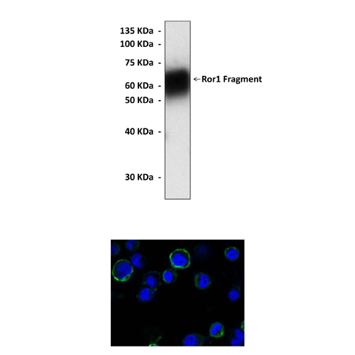
(Click to Enlarge) Top: Western Blot detection of human Ror1 extracellular domain (aa30-406) proteins expressed in HEK293 cell lysate using Ror1 Antibody. Bottom: This antibody stains HEK293 cells expressing human Ror1 extracellular domain (aa30-406) proteins in confocal immunofluorescent analysis (Ror1 Antibody: Green; DRAQ DNA Dye: blue).
Details
Cat.No.: | CP10217 |
Antigen: | Purified recombinant human Ror1 extracellular domain fragments expressed in HEK293 cells. |
Isotype: | Mouse IgG1 |
Species & predicted species cross- reactivity ( ): | Human |
Applications & Suggested starting dilutions:* | WB 1:1000 IP 1:50 IHC n/d ICC 1:200 FACS n/d |
Predicted Molecular Weight of protein: | 135 kDa |
Specificity/Sensitivity: | Detects Ror1 proteins without cross-reactivity with other family members. |
Storage: | Store at -20°C, 4°C for frequent use. Avoid repeated freeze-thaw cycles. |
*Optimal working dilutions must be determined by end user.
Products
| Product | Size | CAT.# | Price | Quantity |
|---|---|---|---|---|
| Mouse Monoclonal Ror1 Antibody: Mouse Monoclonal Ror1 Antibody | Size: 100 ul | CAT.#: CP10217 | Price: $413.00 |