Product Sheet CL0361
Description
BACKGROUND G-CSF belongs to family of colony-stimulating factors, which also includes GM-CSF, IL-3 and M-CSF. It is a potent hematopoietic factor that enhances survival and drives differentiation of myeloid lineage cells, resulting in the generation of neutrophilic granulocytes. Biochemically, G-CSF is an O-glycosylated 19.6 kDa glycoprotein with a pI of 5.5. The biologically active form is a monomer. The analysis of its cDNA has revealed a protein of 207 amino acids containing a hydrophobic secretory signal sequence of 30 amino acids. G-CSF contains 5 cysteine residues, four of which form disulfide bonds (positions 36-42; 64-74). The sugar moiety of G-CSF is not required for full biological activity. G-CSF is secreted by monocytes, macrophages, and neutrophils after cell activation. It is produced also by stromal cells, fibroblasts, and endothelial cells. Epithelial carcinomas, acute myeloid leukemia cells and various tumor cell lines (bladder carcinomas, medulloblastomas), also express this factor. The synthesis of G-CSF can be induced by bacterial endotoxins, TNF, IL1 and GM-CSF. Prostaglandin E2 inhibits the synthesis of G-CSF. In epithelial, endothelial, and fibroblastic cells secretion of G-CSF is induced by IL17.1 G-CSF receptor (G-CSF-R, CD114) is expressed on all cells of the neutrophils and granulocytes lineage. It is expressed also in placenta cells, endothelial cells and various carcinoma cell lines. Human G-CSF is active in murine cells and vice versa. Studies in cell lines have established that conserved tyrosines Tyr704, Tyr729, Tyr744, Tyr764 within the cytoplasmic domain of G-CSF-R contribute significantly to G-CSF-induced proliferation, differentiation, and cell survival. G-CSF-R-mediated activation of JAK tyrosine kinases and signal transducers and activators of transcription (STAT) proteins as well as activation of the ras-MAP kinase route results in induction of gene transcription.2
G-CSF stimulates the proliferation and differentiation of hematopoietic progenitor cells committed to the neutrophils and granulocytes lineage in a dose-dependent manner. At higher concentrations this factor induces the generation of colonies in soft agar cultures containing granulocytes and macrophages. The fully differentiated neutrophilic granulocytes are functionally activated by G-CSF. G-CSF is a mitogen for some human myeloid leukemia cells and also for some carcinoma cell lines. G-CSF synergises with some other cytokines, including GM-CSF and IL4. GM-CSF and G-CSF are required, for example, to develop neutrophilic colonies in vitro. The concerted action of G-CSF and Epo is required to support the growth of mixed colonies of the early erythroid progenitors.2 A combination of IL4 with G-CSF has been shown to lead to synergistic suppression of the growth of some human leukemic cell lines.3
In vitro G-CSF enhances the antibody-dependent cell-mediated cytotoxicity of granulocytes against tumor cells. G-CSF induces the synthesis of receptors for fMLP (Formyl-Met-Leu-Phe) which, in turn, induces the prolonged production of oxygen radicals and the release of arachidonic acid. G-CSF also increases the synthesis of Fc-receptors for IgA. Unlike GM-CSF, G-CSF does not induce the expression of cellular markers such as CD11A and CD11C. The main and most important clinical application of G-CSF is probably the treatment of transient phases of leukopenia following chemotherapy and/or radiotherapy. G-CSF can be used to expand the myeloid cell lineage. It has been shown that the pretreatment with recombinant human G-CSF prior to marrow harvest can improve the graft by increasing the total number of myeloid lineage restricted progenitor cells, resulting in stable but not accelerated myeloid engraftment of autologous marrow.4
REFERENCES
1. Basu, S. et al: Int J Mol Med. 10:3-10, 2002
2. Ward, A.C. et al: Blood 93:113-24, 1999
3. Touw, I.P. & van de Geijn, G.J.: Front Biosci. 12:800-15, 2007
4. Ward, A.C.: Front Biosci.12:608-18, 2007
2. Ward, A.C. et al: Blood 93:113-24, 1999
3. Touw, I.P. & van de Geijn, G.J.: Front Biosci. 12:800-15, 2007
4. Ward, A.C.: Front Biosci.12:608-18, 2007
Products are for research use only. They are not intended for human, animal, or diagnostic applications.
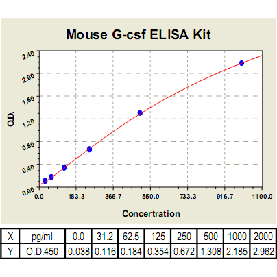
(Click to Enlarge) Granulocyte-Colony Stimulating Factor (G-CSF) ELISA Kit Standard Curve: Using the mouse G-CSF ELISA Kit, O.D. data was graphed against G-CSF protein concentration. The TMB reaction was incubated at 37°C for 14 min.
Details
Cat.No.: | CL0361 |
Target Protein Species: | Mouse |
Range: | 31.2pg/ml-2000pg/ml |
Specificity: | No detectable cross-reactivity with any other cytokine. |
Storage: | Store at 4°C. Use within 6 months. |
ELISA Kits are based on standard sandwich enzyme-linked immunosorbent assay technology. Freshly prepared standards, samples, and solutions are recommended for best results.
Products
| Product | Size | CAT.# | Price | Quantity |
|---|---|---|---|---|
| Mouse G-CSF ELISA Kit: Mouse Granulocyte-Colony Stimulating Factor ELISA Kit | Size: 96 Wells | CAT.#: CL0361 | Price: $619.00 |