Product Sheet CL0369
Description
BACKGROUND Hepatocyte Growth Factor (HGF), also known as Scatter factor (SF), is a multifunctional growth factor that is capable of acting as a potent mitogen, morphogen, motility factor, or angiogenic factor. HGF is a heterodimer of two glycosylated chains, alpha and beta, bound together by a disulfide bond. The molecule is synthesized as single chain precursor devoid of biological activity (pro-HGF). The critical step in pro-HGF activation is a proteolytic cleavage generating the two chain form. This step occurs in the extracellular environment, and is catalyzed by urokinase. Two alternative transcripts originate two HGF variants. One bears a deletion of five amino acids in the alpha chain, and has the same properties of the full-size protein. The other one contains only the first portion of the alpha chain (two kringle HGF). Two kringle HGF binds the HGF receptor, triggers its tyrosine kinase activity and behaves as a partial agonist, inducing motogenesis but not mitogenesis in target cells.1 The HGF receptor is the tyrosine kinase encoded by the c-MET pro-oncogene, a tyrosine kinase receptor. This molecule is an heterodimer of an extracellular alpha chain disulfide linked to a transmembrane beta chain. The cytoplasmic portion of the beta chain contains the catalytic domain and critical sites for the regulation of its kinase activity. HGF binding leads to receptor dimerization and activation of its intrinsic tyrosine kinase, followed by internalization into clathrin-coated vesicles, delivery to sorting endosomes, and degradation via the lysosomal pathway. Phosphorylation of MET at Tyr1230, Tyr1234 and Tyr1235 in the activation loop of the tyrosine kinase domain correlates with increased tyrosine kinase activity. MET activation can lead to autophosphorylation or phosphorylation of downstream intermediates and activation of signaling pathways. Also, Tyr1003 within the juxtamembrane domain recruits c-Cbl (E3-ubiquitin ligase) when phosphorylated. A large number of downstream targets have been defined for MET. As an example, activation of MET with HGF leads to phosphorylation/activation of several pathways involving cell proliferation/survival (ERK1/2, AKT), cell cycle (RB), and cytoskeletal proteins (paxillin, FAK).2 Negative regulation of the receptor kinase activity occurs through distinguishable pathways involving protein kinase C activation or increase in the intracellular Ca2+ concentration. Both lead to the serine phosphorylation of a unique phosphopeptide of the receptor and to a decrease in its kinase activity.3
HGF is secreted by mesenchymal cells and regulates motogenesis, mitogenesis, and morphogenesis of epithelial and endothelial cells.HGF and c-Met are thought to play a key role in normal mesenchymal (site of HGF expression) and epithelial (site of c-Met expression) interactions. HGF has several activities on epithelial cells, including mitogenesis, dissociation of epithelial sheets, stimulation of cell motility, and promotion of matrix invasion. In neoplasia, HGF-c-Met signaling is often aberrant. Constitutively active c-Met mutations underlie hereditary renal carcinoma; c-Met amplification occurs in multiple tumor types and carcinomas frequently possess an autocrine HGF-c-Met loop that correlates with malignant progression.4
HGF is secreted by mesenchymal cells and regulates motogenesis, mitogenesis, and morphogenesis of epithelial and endothelial cells.HGF and c-Met are thought to play a key role in normal mesenchymal (site of HGF expression) and epithelial (site of c-Met expression) interactions. HGF has several activities on epithelial cells, including mitogenesis, dissociation of epithelial sheets, stimulation of cell motility, and promotion of matrix invasion. In neoplasia, HGF-c-Met signaling is often aberrant. Constitutively active c-Met mutations underlie hereditary renal carcinoma; c-Met amplification occurs in multiple tumor types and carcinomas frequently possess an autocrine HGF-c-Met loop that correlates with malignant progression.4
REFERENCES
1. Comoglio, P.M.: EXS. 65:131-65, 1993
2. Faoro, L. et al: J Thorac Oncol. 4(11 Suppl 3): S1064–5, 2009
3. Oka, M. et al: J Invest Dermatol. 128:188-95, 2008
4. Yap, T.A. & de Bono, J.S.: Mol Cancer Ther. 9:1077-9, 2010
2. Faoro, L. et al: J Thorac Oncol. 4(11 Suppl 3): S1064–5, 2009
3. Oka, M. et al: J Invest Dermatol. 128:188-95, 2008
4. Yap, T.A. & de Bono, J.S.: Mol Cancer Ther. 9:1077-9, 2010
Products are for research use only. They are not intended for human, animal, or diagnostic applications.
Details
Cat.No.: | CL0369 |
Target Protein Species: | Human |
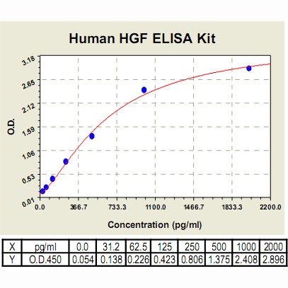
Range: | 31.2 pg/ml – 2000 pg/ml |
Specificity: | NO detectable cross-reactivity with any other cytokine |
Storage: | Store at 4°C. Use within 6 months. |
ELISA Kits are based on standard sandwich enzyme-linked immunosorbent assay technology. Freshly prepared standards, samples, and solutions are recommended for best results.
Products
| Product | Size | CAT.# | Price | Quantity |
|---|---|---|---|---|
| Human HGF ELISA Kit: Human Hepatocyte Growth Factor ELISA Kit | Size: 96 Well | CAT.#: CL0369 | Price: $511.00 |
Resources/Documents
Publications
2017
Guerra, A., W. Rose, P. Hematti and W. Kao. 2017. Minocycline enhances the mesenchymal stromal/stem cell pro-healing phenotype in triple antimicrobial-loaded hydrogels. Acta Biomaterialia, dx.doi.org/10.1016/j.actbio.2017.01.021.

