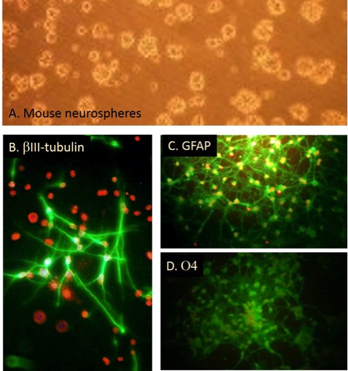
Mouse Neural Stem Cells (MNSC) (A) and Differentiated MNSC immunofluorescently stained with antibodies to βIII-tubulin (B), GFAP (C) and oligodendrocyte marker O4 (D).
Mouse Neural Stem Cells: MNSC
Mouse Neural Stem Cells (MNSC) are multipotent stem cell populations isolated from the developing or adult central nervous system (CNS).
Description
Mouse Neural Stem Cells (MNSC) are multipotent stem cell populations isolated from the developing or adult central nervous system (CNS). As a foundational cell type in developmental neurobiology, these cells possess the unique capacity to proliferate extensively via self-renewal and to undergo multi-lineage differentiation into the primary functional cells of the brain: neurons, astrocytes, and oligodendrocytes. While the terms are sometimes used interchangeably in broad literature, true neural stem cells must be precisely distinguished from more restricted neural progenitor cells (NPCs), or neural precursor cell variants, which possess finite proliferative capacity and a more restricted line of commitment.
Unlike transformed lines, primary MNSC retain a stable diploid karyotype and authentic neurogenic staging profiles. To sustain their undifferentiated, proliferative state in vitro, these fastidious neural cells must be cultured in specialized formulations like an MNSC basal medium or a comprehensive MNSC growth medium kit.
In the living organism, these cells reside within specialized germinal niches of the CNS tissue. During embryonic development, they are located within the ventricular and subventricular zones of the neural tube, actively driving the initial formation of the brain and spinal cord. In the adult mouse brain, restricted neurogenesis persists within two primary neurogenic niches: the subventricular zone (SVZ) lining the lateral ventricles and the subgranular zone (SGZ) of the dentate gyrus within the hippocampus.
In these localized environments, neural stem cells regulate tissue homeostasis by switching their division modes based on structural and biochemical cues; they can divide symmetrically to expand the stem cell pool or asymmetrically to generate a daughter stem cell along with a more restricted neural progenitor cell that migrates to its final anatomical destination to replace damaged neural networks.
The primary biological function of a neural stem cell is to fuel neurogenesis and gliogenesis during development and ongoing tissue maintenance. Guided differentiation can yield functional mature lineages: a differentiated cell can mature into a signaling neuron — marked by the upregulated expression of structural proteins like -III-tubulin (Tuj1) and microtubule-associated protein 2 (MAP2) — or shift toward glial cells, such as GFAP-expressing astrocytes or MBP-positive oligodendrocytes.
In vitro, this multi-lineage potential is classically evaluated by expanding the cells as free-floating cell aggregates known as neurospheres, or as adherent monolayers on specific extracellular matrices, and then tracking the shift from progenitor cell markers (e.g., Nestin, Sox2) to mature lineage-specific proteins. However, investigators must note that traditional neurosphere formation assays select heavily for highly proliferative progenitors and carry inherent limitations, as they are not strictly clonal unless cells are plated at a validated single-cell density to prevent passive aggregation.
In regenerative medicine, molecular biology, and safety pharmacology, MNSC serve as widely used preclinical models that approximate core aspects of mammalian neural repair mechanisms. They serve as a crucial committed-lineage benchmark when evaluating the neural induction efficiency of uncommitted pluripotent stem cells, such as embryonic stem cells (ESCs) or induced pluripotent stem cells (iPSCs), using specific stem cell differentiation kits. However, because they are mouse-derived cells, investigators must account for distinct interspecies differences and functional limitations when projecting outcomes to human neural stem cells, particularly regarding human-specific cortical expansion and unique evolutionary signaling patterns.
Researchers deploy these primary cells to model the pathomechanisms of chronic neurodegenerative disease and traumatic CNS injuries, evaluating the therapeutic potential of cell transplant strategies. In transplant models, scientists examine how effectively transplanted cells survive, integrate, and expand inside damaged host tissue, where they can differentiate into mature, functional neural cells or secrete protective neurotrophic factors within approximately three weeks.
Additionally, in high-throughput drug screening pipelines, MNSC are utilized alongside primary neurons or non-neural controls like bone marrow-derived mesenchymal stem cells to discover neuroprotective compounds and analyze synaptic protein assembly. Because tumorigenicity, hyperplastic cellular proliferation, and uncontrolled host network integration represent very real safety concerns for stem cell therapies, these platforms are vital for conducting rigorous safety assays to evaluate the precise growth kinetics and structural stability of candidate lines before transitioning into translational in vivo models.
Details
| Tissue | Normal healthy mouse brain cerebral cortex | |
|---|---|---|
| QC | No bacteria, yeast, fungi, mycoplasma | |
| Character | β-tubulin III, GFAP & O4 (+) in Diff Med | |
| Bioassay | Form neurospheres in MNSC Gr Med | |
| Cryovial | 2,000,000 MNSC (1st psg) cryopreserved in Freezing Med (040-50) | |
| Kit | Frozen cryovial MNSC (MS820-20f), Grwth Med (R813-250), Neural Stem Cell Dissociation Solution (076-20), 10cm non-TC dish x2 | |
| Proliferating | Shipped in Gr Med, 2nd psg (flasks or plates) | |
| Doublings | At least 2 | |
| Applications | Laboratory research use only (RUO). Not for human, clinical, diagnostic or veterinary use. |
Resources
FAQs
Need More Help?
Visit our comprehensive FAQ page for detailed answers to common questions.
Need More Help?
Visit our comprehensive FAQ page for detailed answers to common questions.
Primary Cell FAQs